

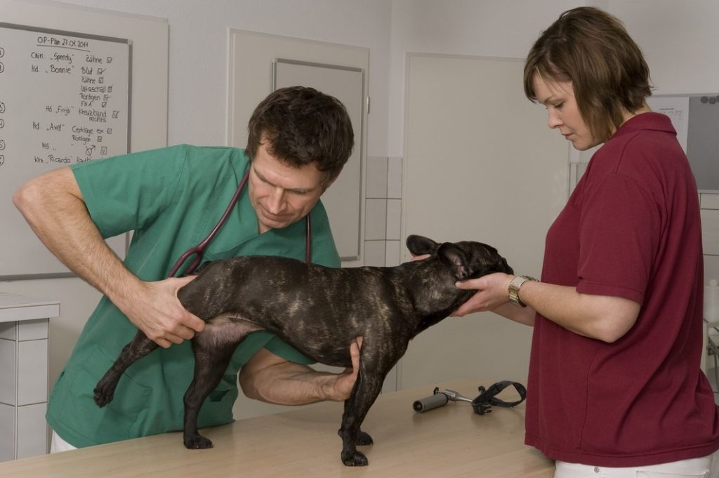
 At Arrowhead, we’ll guide you through the entire process for dealing with your pet’s orthopedic injury — starting at the initial screening and diagnostic stages and continuing through to surgery and post-operative care.
Orthopedic surgery includes both invasive and non-invasive surgeries designed to address issues with the skeletal system and joints, along with associated soft tissues including ligaments, tendons, cartilage and muscles.
Our surgeon, Dr. Ketcher is experienced and successful with her surgeries. Some of the orthopedic surgeries we perform are:
- Fracture repairs
- Hip Surgery
- Femoral head and neck osteotomy (FHNO)
- Knee/MPL/ACL (Human terms)
- Medial patella luxation (MPL)
- Tibial tuberosity advancement (TTA)
- Lateral suture for cranial cruciate ligament repair
CCL Tear
One of the most common disorders affecting canine and feline knees is a cranial cruciate ligament tear, which is analogous to the anterior cruciate ligament (ACL) in people. ACL injuries in humans almost always occur secondary to trauma or excessive force applied to the knee. However, in dogs it is usually a degenerative process that doesn’t necessarily require a traumatic event. Once the ligament is torn, the dog will become lame on the affected limb and may not even be able to bear weight. This may improve slightly over time as the body tries to stabilize the joint, but a significant lameness usually persists. Cranial cruciate injuries can be managed medically, but many dogs will not be able to fully recover without surgical stabilization because the ligament is unable to heal once torn. Diagnosis of the condition is typically made during orthopedic examination and upon X-ray of the knee.
There are many ways to surgically stabilize the knee once the cranial cruciate has been damaged. Two of the more common procedures we offer at Arrowhead include the following: TTA, lateral suture stabilization.
Lateral suture
This is the simplest and most straightforward method of stabilizing the cruciate ligament-deficient knee. The procedure involves the passage of a suture from the back of the femur to the tibial crest in a direction oriented to mimic the absent cranial cruciate ligament. It is used more frequently in smaller dogs and cats. Advantages of the technique include lower risk of complication and expense as the procedure is less invasive than some of the alternatives. Recent publications have shown that this technique is less effective in providing long term comfort and return to function in larger dogs long compared to the next technique we offer for cranial cruciate ligament tear, Tibial Tuberosity Advancement.
TTA
The second surgical technique offered at Arrowhead for cranial cruciate ligament tear is the Tibial Tuberosity Advancement. In this procedure, the front part of the tibia is cut and separated from the rest of the tibia. A special orthopedic spacer is screwed into the space between the two sections of bone to slide the front part of the lower knee forward and up. This moves the patellar ligament (the thick fibrous band that runs on the front of the knee from the top to the bottom of the joint) into better alignment, thereby removing some of the abnormal sliding movement. In some cases, a bone plate is then attached to hold the front section of the tibia in the proper position. By changing the alignment of the patellar ligament, the forces that cause the femur to slip backward when the CCL is torn instead move straight down the tibia, resulting in less instability. TTA offers some benefits over older procedures such as extracapsular repairs, especially for larger or athletic dogs. Dogs undergoing TTA tend to heal faster, resume normal activities quicker, and have a better range of motion in the knee.
MPL
Medial patellar luxation is an orthopedic condition where the kneecap dislocates and becomes misaligned. The condition is both painful and a cause of chronic lameness and arthritis in both dogs and cats. Patellar luxation can also happen along with cruciate ligament ruptures and can even happen genetically in some smaller dog breeds like Yorkshire Terriers. Luxating patellae are often bilateral (both legs affected), but the procedure to correct a luxating patellar is routine with highly successful outcomes.
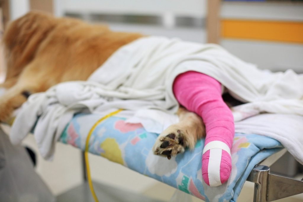
 Fracture repair
Traumatic injuries resulting in bone fractures are common in dogs and cats. Some fractures can be managed with splints or casts. However, more commonly, surgery is indicated to quickly improve the comfort and mobility of your pet. Our surgeon Dr. Ketcher is skilled in several different fracture repair modalities including bone plating/pinning, cerclage wire, and will employ the most practical, safe and effective techniques for your dog or cat.
FHNO
Femoral head and neck osteotomy (FNHO) is a technique by which the femoral head and neck (which comprise the “ball” of the ball-and-socket joint) are surgically removed to alleviate the bone-on-bone pain associated with advanced canine hip dysplasia. It can be completed in any size dog but appears to work best in small dogs and cats whose hip disease is no longer manageable with medical techniques. Advantages of this technique are its low cost; however, it can result in some gait abnormalities and may not eliminate all discomfort in all dogs, especially larger pets.
